MRI of the brain: when necessary, how it goes, contraindications
MRI with the mental abilities are a technique for examining its structure without upsetting the functioning of the organ. It really is employed to examine blood vessels and soft tissues to spot possible injuries and lesions because of strokes.
MRI is completely safe for humans, you can find practically no contraindications. The one limitations matched to the availability of pacemakers and metal implants. A dynamic magnetic field can heat metals in your body or disrupt electronic mechanisms.
Indications for your procedure
MRI of the brain is needed if:
Frequent and severe headaches that cannot be helped by medication.
Dizziness and fainting.
Numbness with the arms and legs, each side weakness inside the limbs.
A clear deterioration in memory.
Regular tinnitus.
Lack of coordination and disorientation wide.
Head trauma.
Progress
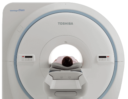
The MRI machine is really a large cylinder when a individual is placed in a supine position. Ahead of the procedure, metal jewelry on the human body, braces, as well as other metal objects are removed. The individual is secured with straps up for grabs to attenuate mobility for the best accurate results.
Special tools are connected to the head that generate and receive radio waves. There's significant noise in the device, so the patient emerges earplugs for maximum comfort.
Research into the result
Inside the resulting image, you can observe veins, neoplasms, dense and soft tissues. The picture is consumed several projections with the desired depth, because of that your doctor can look at the health of any part of the brain. In the scanning process, some images are obtained, because both versions will show a layer-by-layer part of the cerebral tissue. Due to the different contrast the exact same image, every detail might be appreciated.
The pictures show: white matter, cerebral aqueduct, cerebellum, trunk, vascular structures. The tomograph creates images which might be presented in the form of highlighted and eye shadows.
Decryption
When decrypting, a unique interpretation protocol is utilized. The pictures obtained are compared with reference MRI data extracted from a normal brain. To accurately decipher the information received, the specialist has to thoroughly have in mind the physiological and pathological anatomy. It can be obligatory to understand the peculiarities from the operation with the tomograph, that has been useful for the examination.
To get more information about mrt golovnogo mozga check this popular webpage: read

Public Last updated: 2022-01-06 11:43:14 AM