MRI of the brain: when necessary, how it goes, contraindications
MRI in the mental abilities are a technique for examining its structure without interfering with the functioning with the organ. It's accustomed to examine bloodstream and soft tissues to distinguish possible injuries and lesions as a result of strokes.
MRI is completely safe for humans, there are practically no contraindications. The one limitations are matched to the supply of pacemakers and metal implants. An energetic magnetic field can heat metals in the body or disrupt electronic mechanisms.
Indications for your procedure
MRI in the brain is needed if:
Frequent and severe headaches that can not be treated with medication.
Dizziness and fainting.
Numbness in the arms or legs, the look off weakness in the limbs.
A clear, crisp deterioration in memory.
Regular tinnitus.
Loss in coordination and disorientation wide.
Head trauma.
Progress
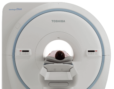
The MRI machine is often a large cylinder in which a individual is put in a supine position. Prior to the procedure, metal jewelry on your body, braces, and also other metal objects are removed. The individual is secured with straps on the table to reduce mobility for the most accurate results.
Special tools are attached to the head that generate and receive radio waves. There exists significant noise within the device, so the patient is provided earplugs for maximum comfort.
Analysis of the result
In the resulting image, you can observe bloodstream, neoplasms, dense and soft tissues. The photo is used several projections with the desired depth, as a result of which the doctor are able to appraise the health of any the main brain. Through the scanning process, a few images are obtained, as both versions will demonstrate a layer-by-layer area of the cerebral tissue. Thanks to the different contrast of the identical image, all the details may be appreciated.
The pictures show: white matter, cerebral aqueduct, cerebellum, trunk, vascular structures. The tomograph creates images that are presented as highlighted and dark areas.
Decryption
When decrypting, a special interpretation protocol is employed. The images obtained are compared with reference MRI data extracted from a healthy brain. To accurately decipher the information received, the specialist must thoroughly understand the physiological and pathological anatomy. It's obligatory to learn the peculiarities of the operation from the tomograph, that was used for the examination.
More information about glavnoe.ua take a look at our site

Public Last updated: 2022-01-06 11:33:36 AM